Mortons Neuroma Solution

Uncategorized

Overview

intermetatarsal neuromaMorton?s Neuroma is a pain condition that affects your feet and toes. If you are suffering from Morton?s Neuroma, a growth of tissue has developed over one of the nerves running from your feet into your toes. This growth can cause inflammation and pain whenever you use your foot. A type of benign tumor, Morton?s Neuroma typically develops in the space between the third and fourth toes, although it can also form between the second and third toes. When you walk, the bones and ligaments in the top of your foot press down on this growth, causing pressure and pain.

Causes

The exact cause of Morton?s neuroma is not known, but the choice of footwear seems be a factor. Wearing high heels (shoes with heels over 2 inches) can put extra pressure on the balls of the feet. Wearing tight or pointed toed shoes may squeeze the toes together or otherwise constrict their movement. For that reason, women are about 8 to 10 times more likely to develop Morton?s neuroma compared with men. People who are born with flat feet, high arches, or an abnormal position of the toes are more prone to developing Morton?s neuroma. This may be due to instability around the toe joints. Certain conditions that develop over time, such as bunions or hammer toes, are also associated with Morton?s neuroma. Some sports that involve running, including tennis and other racquet sports, can also increase the chance of developing Morton?s neuroma due to trauma or injury to the foot.
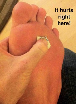
Symptoms

A Morton’s neuroma causes a “burning” sharp pain and numbness on the bottom of the foot in the involved area, and this pain and numbness can radiate to the nearby toes. The pain is usually increased by walking or when the ball of the foot is squeezed together and decreased with massaging. It may force a person to stop walking or to limp from the pain.

Diagnosis

Negative signs include no obvious deformities, erythema, signs of inflammation, or limitation of movement. Direct pressure between the metatarsal heads will replicate the symptoms, as will compression of the forefoot between the finger and thumb so as to compress the transverse arch of the foot. This is referred to as Mulder?s Sign. There are other causes of pain in the forefoot. Too often all forefoot pain is categorized as neuroma. Other conditions to consider are capsulitis, which is an inflammation of ligaments that surrounds two bones, at the level of the joint. In this case, it would be the ligaments that attach the phalanx (bone of the toe) to the metatarsal bone. Inflammation from this condition will put pressure on an otherwise healthy nerve and give neuroma-type symptoms. Additionally, an intermetatarsal bursitis between the third and fourth metatarsal bones will also give neuroma-type symptoms because it too puts pressure on the nerve. Freiberg’s disease, which is an osteochondritis of the metatarsal head, causes pain on weight bearing or compression.

Non Surgical Treatment

Treatment options vary with the severity of each neuroma, and identifying the neuroma early in its development is important to avoid surgical correction. For simple, undeveloped neuromas, a pair of thick-soled shoes with a wide toe box is often adequate treatment to relieve symptoms, allowing the condition to diminish on its own. For more severe conditions, however, additional treatment or surgery may be necessary to remove the tumor. The primary goal of most early treatment regimens is to relieve pressure on areas where a neuroma develops. Your podiatric physician will examine and likely X-ray the affected area and suggest a treatment plan that best suits your individual case. Padding and Taping. Special padding at the ball of the foot may change the abnormal foot function and relieve the symptoms caused by the neuroma. Medication. Anti-inflammatory drugs and cortisone injections can be prescribed to ease acute pain and inflammation caused by the neuroma. Orthotics. Custom shoe inserts made by your podiatrist may be useful in controlling foot function. Orthotics may reduce symptoms and prevent the worsening of the condition.intermetatarsal neuroma

Surgical Treatment

Surgery. This is the last and most permanent course of action. This surgery is used as a last resort as it often comes with a series of side affects including the risk of making the pain worse. This surgery can be performed by Orthopedic surgeons as well as Podiatric surgeons.

What Can Induce Heel Spur

Calcaneal Spur, Heel Spur, Inferior Calcaneal Spur, Posterior Calcaneal Spur

Calcaneal Spur

Overview

The calcaneus is the largest bone in the foot. Pain in the heel region can sometimes be related to Plantar Fasciitis, inflammation of the plantar fascia ligament in the heel area. A heel spur is a hook of bone that forms on the calcaneus where the plantar fascia attaches. Heel spurs can be identified with an X-ray. A heel spur can occur with or without Plantar Fasciitis.

Causes

Fctors that increase the risk of developing heel spurs include a high body mass index (BMI), regular vigorous activity, and intensive training routines or sports. Factors such as these are believed to increase the incidence of repetitive stress injuries that are associated with the formation of heel spurs. When a heel spur forms, extremely sharp pain along with the feeling that a part of the heel is trying to burst through the skin usually occurs. If left untreated, an individual may eventually begin to struggle to perform simple activities such as walking.

Heel Spur

Symptoms

More often than not, heel spurs have no signs or symptoms, and you don?t feel any pain. This is because heel spurs aren?t pointy or sharp pieces of bone, contrary to common belief. Heel spurs don?t cut tissue every time movement occurs; they?re actually deposits of calcium on bone set in place by the body?s normal bone-forming mechanisms. This means they?re smooth and flat, just like all other bones. Because there?s already tissue present at the site of a heel spur, sometimes that area and the surrounding tissue get inflamed, leading to a number of symptoms, such as chronic heel pain that occurs when jogging or walking.

Diagnosis

A Heel Spur diagnosis is made when an X-ray shows a hook of bone protruding from the bottom of the foot at the point where the plantar fascia is attached to the heel bone. The plantar fascia is the thick, connective tissue that runs from the calcaneus (heel bone) to the ball of the foot. This strong and tight tissue helps maintain the arch of the foot. It is also one of the major transmitters of weight across the foot as you walk or run. In other words, tremendous stress is placed on the plantar fascia.

Non Surgical Treatment

Treatment of heel spurs is the same as treatment of plantar fasciitis. Because these problems are related, the treatment is the same. The first step in the treatment of a heel spur is short-term rest and inflammation control. Here are the steps patients should take in order to cure the symptoms of plantar fasciitis and heel spurs. Avoiding the activity that caused the symptoms is the first step in treatment. For example, take a few day off jogging or prolonged standing/walking. Just resting usually helps to eliminate the most severe pain, and will allow the inflammation to begin to cool down. Icing will help to diminish some of the symptoms and control the heel pain. Icing is especially helpful after a sudden flare up of symptoms. Exercises and stretches are designed to relax the tissues that surround the heel bone. Some simple exercises, performed in the morning and evening, often help patients feel better quickly. Many patients will work with a physical therapist, or you can try some simple activities on your own. If you need some help, meet with a therapist for a few sessions to learn a program you can continue on your own.

Surgical Treatment

In some cases, heel spurs are removed by surgery after an X-ray. While the surgery is typically effective, it?s a timely and expensive procedure. Even after surgery, heel spurs can re-form if the patient continues the lifestyle that led to the problem. These reasons are why most people who develop painful heel spurs begin looking for natural remedies for joint and bone pain. Surgery isn?t required to cure a heel spur. In fact, more than 90 percent of people get better with nonsurgical treatments. If nonsurgical methods fail to treat symptoms of heel spurs after 12 months, surgery may be necessary to alleviate pain and restore mobility.

The Right Way To Treat Heel Spur

Calcaneal Spur, Heel Spur, Inferior Calcaneal Spur, Posterior Calcaneal Spur

Calcaneal Spur

Overview

Although a heel spur is often thought to be the source of heel pain, it rarely is. When a patient has plantar fasciitis, the plantar fascia pulls on the bottom of the heel bone. Over time this can cause a spur to form. Heels spurs are a very common x-ray finding, and because the heel spur is buried deep in soft tissue and not truly in a weight bearing area, there is often no history of pain. It is important to note that less than one percent of all heel pain is due to a spur. but frequently caused by the plantar fascia pulling on the heel. Once the plantar fasciitis is properly treated, the heel spur could be a distant memory.

Causes

Heel spurs occur when calcium deposits build up on the underside of the heel bone, a process that usually occurs over a period of many months. Heel spurs are often caused by strains on foot muscles and ligaments, stretching of the plantar fascia, and repeated tearing of the membrane that covers the heel bone. Heel spurs are especially common among athletes whose activities include large amounts of running and jumping. Risk factors for heel spurs include walking gait abnormalities,which place excessive stress on the heel bone, ligaments, and nerves near the heel. Running or jogging, especially on hard surfaces. Poorly fitted or badly worn shoes, especially those lacking appropriate arch support. Excess weight and obesity. Other risk factors associated with plantar fasciitis include increasing age, which decreases plantar fascia flexibility and thins the heel’s protective fat pad. Diabetes. Spending most of the day on one’s feet. Frequent short bursts of physical activity. Having either flat feet or high arches.

Heel Spur

Symptoms

The spur itself is not painful, however, if it is sharp and pointed it can poke into soft tissue surrounding the spur itself. As the bone spur irritates the tissue, inflammation and bruising can occur leading to heel pain. Heel spurs can affect your ability to do your usual work and/or activities, and can also trap and irritate the nerves in your heel area. They can change the way you walk, and can lead to knee, hip and low back injuries. If severe, they may require medical intervention.

Diagnosis

A Diagnosis of Heel Spur Syndrome is a very common reason for having heel pain. Heel pain may be due to other types of conditions such as tendonitis, Haglund’s Deformity, Stress Fracture, Tarsal Tunnel Syndrome, or low back problems. A more common condition in children is Sever’s Disease. The diagnosis is usually made with a combination of x-ray examination and symptoms.

Non Surgical Treatment

Exercise. If you think your pain is exercise-related, change your exercise routine, environment, or foot-ware, and emphasize movements and/or body parts that do not cause pain. Mind/Body. Occasionally foot pain can be related to stress. The body may respond with generalized tension that contributes to pain in many areas, including the feet. Hypnosis and guided imagery are worth exploring if an anatomical problem is not apparent. Supplements. Natural anti-inflammatories can be just as effective as ibuprofen or other over-the-counter pain relievers with fewer side effects. Try one of the following. Ginger (Zingiber officinale) standardized to 5-6% gingerols and 6% shogoals, take one to two 500 mg tablets three to four times daily. Turmeric (Curcuma longa) standardized to 95% curcuminoids, take 400-600 mg three times daily. Also, be sure that you’re wearing well-fitting shoes, the proper shoes for each activity, and that you buy new foot-ware as soon as you notice signs of wear. You can also take the pressure off your heel with a donut-shaped heel cushion or a heel-raising pad placed in your shoe. Acupuncture can also relieve the pain, as can for some sufferers, magnetic shoe inserts, although the evidence behind their effectiveness is not conclusive. Osteopathic or chiropractic manipulation can help with soft tissue pain in and around the feet.

Surgical Treatment

Surgery to correct for heel spur syndrome is a common procedure which releases plantar fascia partially from its attachment to the calcaneous (heel bone). This part of the surgery is called a plantar fasciotomy due to the fact the fascia is cut. This is most often done through an open procedure as any heel spur or bursa can be removed at the same time. If the spur is not removed during the surgery, it will probably be just as successful, as the large spur is not the true problem. Some physicians use an endoscopic approach (EPF) where a small camera aids the physician during surgery with typically smaller incisions on each side of your foot.

Bursitis Of The Foot Surgical Procedures

Bursitis, Foot Pain

Overview

A lesser known type of heel pain is a condition called Bursitis of the Heel. A bursa is a fluid-filled sac that cushions the muscles, tendons and bones in our joints. It helps keep them from rubbing against each other and reduces friction in the areas around the joints. Bursitis is Latin for inflammation of the bursa. Repeated movement and pressure on the bursa can cause it to swell and become inflamed. Trauma, infection or crystal deposits can also cause Bursitis. The joints that are usually affected by bursitis are the large joints such as the shoulder, hip and knee but in some cases also the back of the heel.

Causes

Causes of bursitis can be from any form of friction between bone and the soft tissues. The most common cause is due to abnormal pronation.

Symptoms

Your heel may feel more sensitive to the cold and ache in cold and damp weather due to impaired circulation. These symptoms are often the result of failure to treat the injury properly from the outset and overicing.

Diagnosis

Your doctor will check for bursitis by asking questions about your past health and recent activities and by examining the area. If your symptoms are severe or get worse even after treatment, you may need other tests. Your doctor may drain fluid from the bursa through a needle (aspiration) and test it for infection. Or you may need X-rays, an MRI, or an ultrasound.

Non Surgical Treatment

One of the most effective treatments for infracalcaneal bursitis is to temporarily avoid weight-bearing activities that put stress or strain on your heel bone. PRICE (protection, rest, hot/cold contrast compresses, compression, and elevation) is another good acute management technique for this foot problem. Changing your footwear is an effective long-term prevention and treatment tool for infracalcaneal bursitis. Footwear that possess a flat support base, a sufficiently wide toe box to accommodate natural toe splay, and a flexible sole are best for preventing and managing infracalcaneal bursitis. An integrated approach to this problem usually involves the use of padded socks that help reduce pressure, friction, and inflammation in your affected area. Natural anti-inflammatory agents can also be helpful in quelling inflammation, reducing pain, and improving treatment times for infracalcaneal bursitis. In rare cases, more aggressive treatment methods may be required, such as cortisone injections or surgery to drain the bursal sac.

Surgical Treatment

Surgery. Though rare, particularly challenging cases of retrocalcaneal bursitis might warrant a bursectomy, in which the troublesome bursa is removed from the back of the ankle. Surgery can be effective, but operating on this boney area can cause complications, such as trouble with skin healing at the incision site. In addition to removing the bursa, a doctor may use the surgery to treat another condition associated with the retrocalcaneal bursitis. For example, a surgeon may remove a sliver of bone from the back of the heel to alter foot mechanics and reduce future friction. Any bone spurs located where the Achilles attaches to the heel may also be removed. Regardless of the conservative treatment that is provided, it is important to wait until all pain and swelling around the back of the heel is gone before resuming activities. This may take several weeks. Once symptoms are gone, a patient may make a gradual return to his or her activity level before their bursitis symptoms began. Returning to activities that cause friction or stress on the bursa before it is healed will likely cause bursitis symptoms to flare up again.

Prevention

You can avoid the situation all together if you stop activity as soon as you see, and feel, the signs. Many runners attempt to push through pain, but ignoring symptoms only leads to more problems. It?s better to take some time off right away than to end up taking far more time off later. Runners aren?t the only ones at risk. The condition can happen to any type of athlete of any age. For all you women out there who love to wear high-heels-you?re at a greater risk as well. Plus, anyone whose shoes are too tight can end up with calcaneal bursitis, so make sure your footwear fits. If the outside of your heel and ankle hurts, calcaneal bursitis could be to blame. Get it checked out.

Non Surgical Hammer Toe Correction

Hammer Toe

Hammer ToeOverview

A hammertoes is a misshapen toe. The middle joint of the toe bends up in a way that makes the toe look like it is forming an upside-down V. The bent joint may rub the top of your shoe. Hammertoes can develop on any toe, but they usually happen in the second toe. Claw toes or mallet toes look a lot like hammertoes, but a different joint in the toe is bent. Hammertoes usually are not painful at first. When they begin, they can be pushed down to the correct position. These are called flexible hammertoes. After a while, they will not go back to their normal position, even if pushed with the fingers. These are called rigid hammertoes.

Causes

Though hammer toes are principally hereditary, several other factors can contribute to the deformity. Most prevalent is an imbalance of the muscles and tendons that control the motion of the toe. When the tendon that pulls the toe upward is not as strong as the one that pulls it downward there is a disparity of power. This forces the toe to buckle and gradually become deformed. If the it persists, the toe can become rigid and harder to correct.

HammertoeSymptoms

If you have any of these symptoms, do not assume it is due to hammer toe. Talk to your doctor about symptoms such as a toe that curls down, corns on the top of a toe, calluses on the sole of the foot or bottom of the toe, pain in the middle joint of a toe, discomfort on the top of a toe, difficulty finding any shoes that fit comfortably, cramping in a toe, and sometimes also the foot and leg, difficult or painful motion of a toe joint, pain in the ball of the foot or at the base of a toe.

Diagnosis

Your healthcare provider will examine your foot, checking for redness, swelling, corns, and calluses. Your provider will also measure the flexibility of your toes and test how much feeling you have in your toes. You may have blood tests to check for arthritis, diabetes, and infection.

Non Surgical Treatment

In the earlier stages of hammer toe, when the toes can still be manually straightened, then conservative treatment is appropriate. This means wearing shoes which are a half size bigger than normal and which are not narrow around the toes. Exercises to stretch the toes out and Hammer toes strengthen the muscles under the foot which balances the tightness of the top tendons are important. Padding or corn plasters can be used to ease the discomfort of any associated corns and calluses.

Surgical Treatment

Your podiatrist may recommend a surgical procedure if your hammertoes are not helped by the conservative care methods listed above. Surgery for hammertoes is performed to help straighten your crooked toe. Your surgery will be performed in your podiatrist?s office or at a hospital, depending on the severity of your hammertoe. A metal pin is sometimes used to help your affected toe maintain its straight position during your recovery.

Bunions Causes Symptoms And Treatments

Uncategorized

Overview
Bunions
In constrictive shoes, the big toe is forced to bend toward the second toe and the first joint of the big toe is moved out of place. To compensate for the realignment, the outside of the joint is increased in size. Tendons then begin to pull the toe into an abnormal position. Over time the change in position becomes painful and permanent. The change in position also causes the mechanics of the toes and foot to be affected. The joint at the base of the big toe carries a lot of weight when walking or running. In a normally shaped foot the position of the big toe helps create a wide base of support and stability. A foot that has had the big toe bent toward the second toe will tend to roll inward. This abnormal pronation, along with the ill-fitting shoes will make the Bunion even worse. If a person has a foot anatomy that is prone to Bunions, wearing footwear with a too-narrow toe box will accelerate the development of a Bunion. Wearing footwear with a wide toe box may help prevent or at least delay the development of Bunions.

Causes
Bunions are most often caused by faulty mechanics of the foot. The deformity runs in families, but it is the foot type that is hereditary, not the bunion.Certain foot types make a person prone to developing a bunion. Injury can be a cause, especially if just one foot is involved. Although wearing shoes that crowd the toes won?t actually cause bunions in the first place, it can make the deformity progressively worse. That means you may expererince symptoms sooner.
SymptomsSymptoms, which occur at the site of the bunion, may include pain or soreness, inflammation and redness, a burning sensation, possible numbness. Symptoms occur most often when wearing shoes that crowd the toes, such as shoes with a tight toe box or high heels. This may explain why women are more likely to have symptoms than men. In addition, spending long periods of time on your feet can aggravate the symptoms of bunions.

Diagnosis
Your doctor is very likely to be able to diagnose your bunion simply by examining your foot. Even before that, he or she will probably ask about your family and personal medical history and evaluate the types of shoes you wear. You’ll be asked about your symptoms, when they started and when they occur. You may also be asked to flex your toe so that your doctor can get an idea of your range of motion. He or she may order x-rays in order to determine the extent of your deformity.

Non Surgical Treatment
Initial treatment of bunions may include wearing comfortable, well-fitting footwear (particularly shoes that conform to the shape of the foot and do not cause pressure areas) or the use of splints and orthotics (special shoe inserts shaped to your feet) to reposition the big toe. For bunions caused by arthritis, medications may help reduce pain and swelling. If nonsurgical treatment fails, your doctor may suggest surgery, which resolves the problem in nearly all persons. The goal of surgery is to relieve pain and correct as much deformity as possible. The surgery is not cosmetic and is not meant to improve the appearance of the foot. Other related procedures that may be used to help diagnose foot disorders include X-rays of the bone and foot.
Bunion Pain

Surgical Treatment
This involves surgically correcting the deformity and can involve a variety of different methods. However, outcomes can be variable. This is very dependant of the amount of damage to the joint and the procedure used to correct it. Removal of the bunion is performed using different methods that are out of the scope of this article. Unfortunately, bunions can recur following surgery, and even if it surgery is successful, around 30% of patients still report existing difficulties.

Over-Pronation Of The Feet Suffering

Overpronation

Overview

Over-pronation, or flat feet, is a common biomechanical problem that occurs in the walking process when a person’s arch collapses upon weight bearing. This motion can cause extreme stress or inflammation on the plantar fascia, potentially causing severe discomfort and leading to other foot problems.Over Pronation

Causes

Over-pronation may happen because the tissue that attaches to your foot bones is loose. You may be born with this problem or it may result from injuries or overuse, like from too much running.

Symptoms

Overpronation causes alterations in proper muscle recruitment patterns leading to tightness in the outside of the ankle (lateral gastrocnemius, soleus, and peroneals). This tightness can lead to weakness in the opposing muscles such as the medial gastrocnemius, anterior tibialis, and posterior tibialis. If these muscles are weak, they will not be able to keep the knee in proper alignment, causing the valgus position. All this tightness and weakness can cause pain within the ankle, calf, and knee region. And it can send imbalance and pain all the way up to the upper back, if deep core strength is lacking and can’t hold the pelvis in neutral.

Diagnosis

When sitting, an over-pronating foot appears quite normal, i.e. showing a normal arch with room under the underside of the foot. The moment you get up and put weight on your feet the situation changes: the arches lower and the ankle slightly turns inwards. When you walk or run more weight is placed on the feet compared to standing and over-pronation will become more evident. When walking barefoot on tiles or timber floors over-pronation is more visible, compared to walking on carpet or grass.Overpronation

Non Surgical Treatment

Fortunately, there are simple things you can do to cure and correct your overpronation issues. Certain exercises help. Pull your toes back using a rolled up towel. Roll your feet over a golf or tennis ball for a minute. And do calf raises by standing up and lifting up on your toes. These all help reposition the foot and strengthen the muscles and tendons necessary for proper support. Beyond that, simple adjustments to footwear will help immensely.

Prevention

Wearing the proper footwear plays a key role as a natural way to help pronation. Pronated feet need shoes that fit well, provide stability, contain supportive cushioning, are comfortable and allow enough room for your foot to move without causing pain or discomfort. Putting special inner heel wedges, known as orthotics, into your shoes can support a flatfoot while lowering risks of developing tendinitis, according to the American Academy of Orthopaedic Surgeons. More extensive cases may require specially fitted orthopaedic shoes that support the arches.

Physiotherapy And Severs Disease

Severs Disease

Overview

The muscle group at the back of the lower leg is commonly called the calf. The calf comprises of 2 major muscles (known as the gastrocnemius and soleus) both of which insert into the heel bone via the Achilles tendon. In people who have not yet reached skeletal maturity, a growth plate exists where the Achilles tendon inserts into the heel bone. This growth plate is primarily comprised of cartilage. Every time the calf contracts, it pulls on the Achilles tendon which in turn pulls on the heel’s growth plate. When this tension is too forceful or repetitive, irritation to the growth plate may occur resulting in pain and sometimes an increased bony prominence at the back of the heel. This condition is called Severs disease. Severs disease is typically seen in children or adolescents during periods of rapid growth. This is because muscles and tendons become tighter as bones become longer. As a result, more tension is placed on the heel’s growth plate.

Causes

Sever’s disease also can result from standing too long, which puts constant pressure on the heel. Poor-fitting shoes can contribute to the condition by not providing enough support or padding for the feet or by rubbing against the back of the heel. Although Sever’s disease can occur in any child, these conditions increase the chances of it happening, pronated foot (a foot that rolls in at the ankle when walking), which causes tightness and twisting of the Achilles tendon, thus increasing its pull on the heel’s growth plate, flat or high arch, which affects the angle of the heel within the foot, causing tightness and shortening of the Achilles tendon, short leg syndrome (one leg is shorter than the other), which causes the foot on the short leg to bend downward to reach the ground, pulling on the Achilles tendon, overweight or obesity, which puts weight-related pressure on the growth plate.

Symptoms

Patients with Severs disease typically experience pain that develops gradually in the back of the heel or Achilles region. In less severe cases, patients may only experience an ache or stiffness in the heel that increases with rest (especially at night or first thing in the morning). This typically occurs following activities which require strong or repetitive contraction of the calf muscles, such as running (especially uphill) or during sports involving running, jumping or hopping. The pain associated with this condition may also warm up with activity in the initial stages of the condition. As the condition progresses, patients may experience symptoms that increase during activity and affect performance. Pain may also increase when performing a calf stretch or heel raise (i.e. rising up onto tip toes). In severe cases, patients may walk with a limp, have difficulty putting their heel down, or be unable to weight bear on the affected leg. Pain may also increase on firmly touching the affected region and occasionally a bony lump may be palpable or visible at the back of the heel. This condition typically presents gradually overtime and can affect either one or both lower limbs.

Diagnosis

Low-grade inflammation of the calcaneal apophysis cannot be seen on x-ray. Therefore, although x-rays are often done to rule out bony injuries in children with Sever’s disease these x-rays are usually normal. Advanced Sever’s disease can be seen on x-ray but usually the problem is treated before it reaches this point. Other diagnostic tests, such as bone scans or MRI’s, are not usually required in typical cases of Sever’s disease. These, or other tests, may be required to rule out other conditions, such as stress fractures of the calcaneus or other bony abnormalities that can mimic Severs disease.

Non Surgical Treatment

Ice the heel(s) well after exercise (until the area is cold and numb!) Stretch hamstring and calf muscles 2-3 times daily (exercises below) REST when pain becomes persistent or moderate (even if it means skipping games or practices.) Anti-inflammatory medication such as ibuprofen. If symptoms persist, your child may need to see a physical therapist for additional exercises, and/or an orthopedist for othotics or temporary casting/crutches if pain is severe. Sever?s disease is self-recovering, meaning that it will go away on its own when the heels are rested or when the bone is through growing. The condition is not expected to create any long-term disability, and expected to subside in 2-8 weeks. However, pain can recur, for example at the start of a new sports season, several times if it is not taken care of.

Do You Really Understand Heel Pains?

Heel Pain

Overview

Feet Pain

Your foot is made up of 26 bones, 33 joints, and more than 100 tendons. The heel is the largest bone in your foot. If you overuse or injure your heel, you may experience Heel Pain. This can range from mild to disabling. In many cases, if you have heel pain, you will need a doctor to diagnose the cause.

Causes

Near the inflamed plantar fascia attachment, but not in it, some extra bone may form, producing a small “spur”. In fact, it is a shelf of bone, not a sharp spur. These “heel spurs” are commoner in people with plantar fascitis, but they can be found in people with no heel pain. The heel spur is caused by the same process as the heel pain, but the spur is not itself the cause of the pain.

Symptoms

Symptoms of plantar fasciitis vary, but the classic symptom is pain after rest–when you first get out of bed in the morning, or when you get up after sitting down for a while during the day. The pain usually diminishes after a few minutes of walking, sometimes even disappearing, but the pain is commonly felt again the longer you’re on the foot. Fasciitis can be aggravated by shoes that lack appropriate support, especially in the arch area, and by the chronic irritation of long-periods of standing, especially on concrete, by being overweight. It doesn’t help that fascia doesn’t heal particularly quickly because it has relatively poor circulation (which is why it’s white in colour).

Diagnosis

A biomechanical exam by your podiatrist will help reveal these abnormalities and in turn resolve the cause of plantar fasciitis. By addressing this cause, the patient can be offered a podiatric long-term solution to his problem.

Non Surgical Treatment

Recommended treatments, heel Spurs: cushioning for the heel is of little value. Your chiropodist/podiatrist may initially apply padding and strapping to alter the direction of stretch of the ligament. This is often successful at reducing the tenderness in the short term. Your chiropodist/podiatrist may suggest a course of deep heat therapy to stimulate the healing processes, allowing damage to respond and heal faster. In the long term, your chiropodist/podiatrist may prescribe special insoles (orthoses) to help the feet to function more effectively, thereby reducing strain on the ligaments and making any recurrence less likely. If pain from heel spurs continues, you may be referred to your GP who can prescribe an oral non-steroidal anti-inflammatory. Alternatively, localised hydrocortisone injection treatment may be given by your GP or an appropriate chiropodist/podiatrist. If pain persists, surgery may be considered. Heel Bursitis: in most cases, attention to the cause of any rubbing, and appropriate padding and strapping by your chiropodist/podiatrist will allow the inflammation to settle. If infection is present, your chiropodist/podiatrist will refer you to your GP for antibiotics. Heel Bumps: adjustments to footwear is often enough to make them comfortable. A leather heel counter and wearing boots may help. However, if pain persists, surgery may be necessary.

Surgical Treatment

Only a relatively few cases of heel pain require surgery. If required, surgery is usually for the removal of a spur, but also may involve release of the plantar fascia, removal of a bursa, or a removal of a neuroma or other soft-tissue growth.

Prevention

Pain Of The Heel

Prevention of heel pain involves reducing the stress on that part of the body. Tips include. Barefeet, when on hard ground make sure you are wearing shoes. Bodyweight, if you are overweight there is more stress on the heels when you walk or run. Try to lose weight. Footwear, footwear that has material which can absorb some of the stress placed on the heel may help protect it. Examples include heel pads. Make sure your shoes fit properly and do not have worn down heels or soles. If you notice a link between a particular pair of shoes and heel pain, stop wearing them. Rest, if you are especially susceptible to heel pain, try to spend more time resting and less time on your feet. It is best to discuss this point with a specialized health care professional. Sports, warm up properly before engaging in activities that may place lots of stress on the heels. Make sure you have proper sports shoes for your task.